SCI论文(www.lunwensci.com):
摘要:胰腺癌因诊断不及时而预后极差。因而早期诊断对于改善预后至关重要。临床证据提示胰腺癌的患者应接受正规诊断,而可能罹患胰腺癌的高危人群(例如:急性加重的糖尿病、急性反复性胰腺炎、慢性胰腺炎患者等)应接受随访观察。通过影像学、肿瘤标记物、家族遗传史调查等方式筛选出存在高风险的病例有助于提高胰腺癌的早期诊断率。超声、CT、MRI及ERCP对于早期诊断胰腺癌的能力有限,而内镜检查如超声内镜、内镜下细针穿刺和内镜下采集胰液进行细胞学检查越来越受到重视。展望未来,应该寻找新的肿瘤标记物,与影像学手段相结合,提高胰腺癌的早期诊断率,改善预后。
关键词:胰腺癌;早期诊断
本文引用格式:孔祥耀,潘耀振.胰腺癌早期诊断的最新关注点[J].世界最新医学信息文摘,2019,19(98):80-82.
The Latest Focus of Early Diagnosis of Pancreatic Cancer
KONG Xiang-yao,PAN Yao-zhen*
(Guizhou Medical University,Guiyang Guizhou)
ABSTRACT:The prognosis of pancreatic cancer is very poor because of the untimely diagnosis.Therefore,early diagnosis of pancreatic cancer is very important for improving prognosis.Clinical evidence suggests that patients with pancreatic cancer should receive formal diagnosis,and those at high risk for pancreatic cancer(such as acute exacerbated diabetes,acute recurrent pancreatitis,chronic pancreatitis,etc.)should be followed up.Screening high-risk cases through imaging,tumor markers,and family genetic history surveys can help improve the early diagnosis rate of pancreatic cancer.Ultrasound,CT,MRI and ERCP are limited in the early diagnosis of pancreatic cancer,while endoscopy,such as endoscopic ultrasonography,fine needle aspiration and endoscopic pancreatic juice collection for cytological examination,has attracted more and more attention.Looking forward to the future,we should look for new tumor markers and combine them with imaging methods to improve the early diagnosis rate and prognosis of pancreatic cancer.
KEY WORDS:Pancreatic Cancer;Early Diagnosis
1介绍
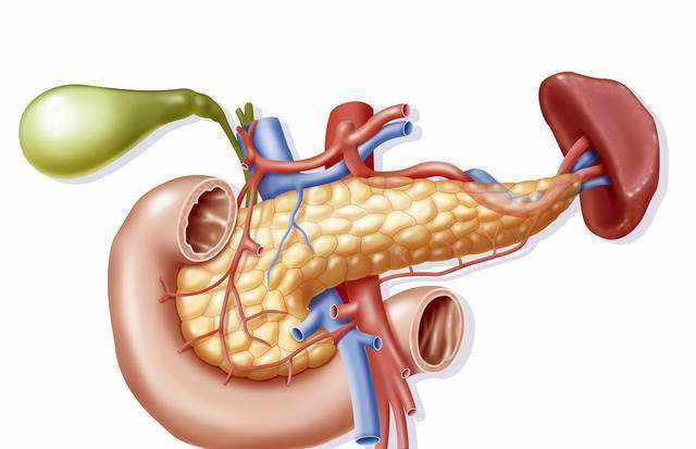
胰腺癌是一种预后极差的恶性肿瘤,发病率逐年升高,预计将在2030年成为全球排名第二的癌症相关死亡原因[1-3]。胰腺导管腺癌是胰腺癌中最常见的类型。多数患者在就诊时,已经出现局部晚期或远处转移,只有少部分患者适合以治疗为目的的外科切除[3]。胰腺癌晚期患者的预后普遍较差,很大程度上归因于胰腺癌早期诊断十分困难[4]。为此对于这些患者,最重要的是尽早接受正规的医学诊断。然而,通过侵入性手术获取组织虽可以得到最准确的诊断但常得不偿失。目前诊断金标准是超声引导的内窥镜细针抽吸(US-FNA),可以进行细胞学分析及诊断,但其缺点是并不能保证每次都能得到完整的分析结果[5-6]。根据日本胰腺协会数据分析,肿瘤直径≤10mm的患者五年生存率达到了80.4%[7],提示胰腺癌早期诊断在提高胰腺癌预后上极其重要。出于以上原因,迫切需要新的技术手段或肿瘤学标志物来协助诊断、分期和指导临床治疗决策。
2胰腺癌早期诊断的最佳时机
研究提示,之所以胰腺癌的预后极差,是因为约有90%的胰腺癌患者在Ⅲ期甚至Ⅳ期时才得到确诊[8]。Haeno等通过对228例胰腺癌患者数据(包括101例尸检)进行了数学构架模型分析,结果提示所有病例均在应该被诊断的黄金时期被漏诊。该数学构架模型提示当原发肿瘤直径≤10mm时,患者约有28%的几率未得到正确诊断。当原发肿瘤直径增加至20mm至30mm时,漏诊率分别增加至73%至94%[9]。此研究提示,当胰腺癌肿瘤直径≤10mm且恶性程度较低时,可被定义为早期胰腺癌,预后通常较好。Hruban等首次报道了对胰腺癌癌前病变胰腺上皮内瘤变病例进行统计学分析后总结出一套遗传进展模型[10],根据细胞异型程度不同将胰腺上皮内瘤变划分为PanIN-1、PanIN-2及PanIN-3三大类。研究表明PanIN-1中普遍存在K-ras异常,PanIN-2中普遍存在p16异常下调,PanIN-3中则普遍存在TP53和SMAD4异常下调[11-16]。近期有一项研究通过计算机对一定数量的尸体解剖结果进行数据分析,建立了一种胰腺癌的进展期生存数据模型[17-18]。该数据模型表明大部分胰腺癌患者在这个生存时间的最后阶段才得到确诊,表明在胰腺癌的发展历程中,诊断较晚往往提示着极差的预后。而在早期胰腺癌逐步发展至晚期胰腺癌,平均需要约2至3年的时间,因此,胰腺癌留给我们的最佳诊断时间窗约为2-3年。尽早的诊断直接关系到胰腺癌的预后。
3胰腺癌的危险因子及癌前病变
2013年,日本胰腺协会(JPS)基于循证医学发布了胰腺癌临床指南,对胰腺癌的诊断及治疗提供了标准[19]。指南认为相关危险因素会加快胰腺癌的疾病进展,归纳总结胰腺癌相关危险因素包含家族遗传史、伴随疾病、生活习惯三大方面。其中家族遗传史包括:(1)亲属患有胰腺癌;(2)患有遗传性胰腺癌综合征;伴随疾病包括肥胖、糖尿病、慢性胰腺炎、遗传性胰腺炎、胰腺囊肿及导管内乳头状粘液瘤;生活习惯则包括吸烟和酗酒。当患者有超过1项危险因子时,建议完善更深入的检查来明确是否患有胰腺癌。报道称部分胰腺导管内乳头状瘤病例在随访过程中会转变为胰腺癌[20-21]。此外,一部分回顾性研究表明胰腺癌患者中合并患有导管内乳头状瘤的概率可达11.2%。日本学者研究报告称,在349例分支管道导管内乳头状瘤病例随访过程中,检测到7例转变成了胰腺癌[22],并称导管内乳头状瘤的诊断比胰腺癌更早,这有助于将这部分病人提早筛选出来予以随访观察处理[23]。
一项关于慢性胰腺炎的前瞻性研究表明,在慢性胰腺炎5年、10年、20年的观察中,胰腺癌的发生率分别为1.1%,1.8%和4.0%[24-25]。
统计显示,约有24%的胰腺癌患者合并患有糖尿病[26]。一项荟萃分析提示,糖尿病患者罹患胰腺癌的风险是非糖尿病患者的两倍[27]。对于糖尿病患者,尤其是当年龄超过50岁时,约有1%的患者在3年内罹患胰腺癌[28]。值得注意的是,糖尿病急性加重有可能是胰腺癌的征象,当糖尿病患者出现无法解释的病情急性加重时,应当考虑到这一点,以免耽误治疗时机。
家族性遗传性胰腺炎是胰腺炎一种罕见类型,通常起病年龄较早,有家族聚集性,胰腺癌的发病率较高。家族性遗传性胰腺炎的诊断通常需满足以下条件:(1)有两个或两个以上的家族成员罹患急性复发性胰腺炎或慢性胰腺炎;(2)家族成员患者通常无酗酒史;(3)至少有一名年轻家族成员患者(40岁以下)患过胰腺炎。但如果患者基因PRSS1存在p.R122H或p.N291突变,则可确立遗传性胰腺炎的诊断,且无需满足以上条件。遗传性胰腺炎的患者,罹患胰腺癌的相对危险度为50,在50岁、60岁和75岁遗传性胰腺炎患者中,引起胰腺癌的比例分别为10%、18.7%和53.5%[29]。据报道,在家族性胰腺癌患者病例中,乳腺癌基因2(BRCA2)、乳腺癌易感基因2(PALB2)和共济失调性毛细血管扩张症较为常见[30-32]。据统计,在胰腺癌患者中,约有5%至10%的患者有胰腺癌家族史。根据美国国家家族性胰腺癌登记处(NFPTR)统计数据显示,有胰腺癌家族史的人群罹患胰腺癌的机率较正常人高6.8倍。近期研究表明,一些遗传相关的危险因子、综合征与胰腺癌的关系日益密切。例如,遗传性胰腺炎(spink1突变),遗传性乳腺卵巢综合征(BRCA1和BRCA2突变,布加迪综合征(STK11/LKB1突变),家族性非典型多发性葡萄胎综合征(CDKN2A(p16)突变),Lynch综合征(MLH1,MSH2,MSH6,或PMS2缺失)与胰腺癌密切相关[33-35]。
以上结论提示患有胰腺癌癌前病变的胰腺导管内乳头状瘤、胰腺囊肿、慢性胰腺炎的患者,即使无症状,仍应定期进行6个月的影像检查,接受更加细致、规范的随访观察,以便更早进行医学干预,得到更理想的预后。在临床上值得关注的是,当检查胰腺炎患者时,应考虑到其有无遗传性胰腺炎家族史。针对有遗传危险因子和综合征的人群,一些研究表明应用核磁共振超声或超声内镜(EUS)对诊断胰腺癌和具有恶性倾向的癌前病变具有一定潜力。
4肿瘤标记物
迄今为止,临床上唯一广泛使用的是测定碳水化合物抗原19-9(CA19-9);其他血清标志物例如癌胚抗原(CEA)和人胰腺癌细胞抗原-2(DUPAN-2)在临床上也有广泛应用。然而,由于特异性有限,它们一般仅用作诊断和肿瘤复发的检测指标,而用于胰腺癌的早期诊断效果并不令人满意[36-37]。值得注意的是,最近一些研究通过从血液中获取循环肿瘤细胞(CTC)、循环游离DNA(CFDNA)或RNA、外显子体和分泌体的方式,从中获取信息用于分析,所有这些检验方法都属于体液检查方法[38-40]。近些年,一些利用血样本或体液样本,从生理学、免疫学和遗传学的角度挖掘出的有潜力的肿瘤标记物亦陆续被报道[41]。有研究称,基于血清代谢组学的诊断模型和唾液转录组学瘤标相比传统指标对于可切除的胰腺癌的诊断有更高的准确性[42]。Fukutake等报道,通过对胰腺癌血浆游离氨基酸与健康对照组相比较,发现两者在组成成分上有显著差异,这对筛查和诊断胰腺癌而言将是一个崭新的,有潜力的研究方向[43]。另有一些研究报道称,利用循环血液中外泌体的一组Micro-RNA作为胰腺癌的诊断标记物表现出很大潜力。Yu等通过利用QR-PCR对胰腺高级别上皮内瘤变(PanIN-3)患者中采集的胰液进行分析,发现700例患者中有35例存在mi-RNA的表达异常[44]。Kojima等报道了利用10个重要的miRNAs协同诊断胰腺癌,此项研究表明利用mi-RNA对于筛选出可切除的胰腺癌有一定效力和临床意义[45]。
5影像学检查
约翰霍普金斯大学一项大型胰腺癌筛查项目中,五个医疗中心通过CT、MRI和EUS检查从可能罹患胰腺癌的高危人群中筛查出共216例疑似胰腺癌的病例。其中,有92例(约43%)病例最终证实为胰腺包块或胰腺导管扩张。CT、MRI和EUS检出率分别为11%,33.3%和43.6%[46]。另一项为期5年的德国研究通过EUS,MRCP和实验室检查筛查出了共76例可能罹患胰腺癌的高危病例。这些研究将胰腺高级上皮内瘤变(PanIN-3)的阳性检出率提高到了1.3%[47]。以上研究数据提示,对于可能罹患胰腺癌的高危人群进行定期影像学筛查常可检测出胰腺微小囊性病变,包括癌前病变。
超声应作为胰腺癌早期诊断首选的影像学检查方式。有研究称应用超声检测到主胰管的微小扩张和胰腺囊肿是胰腺癌早期较为重要的预测指标之一。Tanaka等通过超声检查从1058例高危人群(通过超声检查提示存在胰管扩张或胰腺囊肿的人群)的随访观察中诊断出12例胰腺癌[48],提示超声检查对于尽早筛选胰腺癌患者有一定价值,且超声检查方便、廉价,应提倡在基层医院广泛展开使用,但考虑到超声检查主观性较强,受超声科医生水平限制,超声医师应得到更加专业的培训。
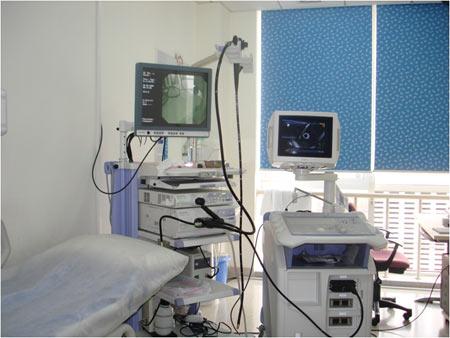
超声内镜是一种侵入性检查,然而,其诊断胰腺癌的优越性已被广泛报道[49]。尤其是相对于MDCT(多排螺旋CT)而言[50]。MRI在发现胰腺囊性病变上有巨大优势,而超声内镜在检测实体肿瘤上亦有不俗表现[51]。Yasuda等回顾性分析了132例可能罹患胰腺癌的高危人群,以上患者通过CT检查并未检测到包块,但通过超声内镜检查从以上病例中检测出3例肿块直径小于10mm的胰腺癌患者[52],提示超声内镜在诊断胰腺癌方面相对CT仍有一定优势,CT无法检测到包块的原因可能与CT扫描的厚度直接相关,而超声内镜通过天然腔隙可近距离扫描病变组织,这是超声内镜独特的优势所在。Kamata等通过每半年一次EUS、每年一次超声检查以及CT、MRI分别对102例可能罹患胰腺癌的高危人群进行长期随访观察,11例胰腺癌伴胰腺导管内乳头状粘液肿瘤的患者在首次检查时通过EUS诊断出来,7例胰腺癌伴胰腺导管内乳头状粘液肿瘤患者在随访中得到确诊[53]。以上研究提示EUS在胰腺癌的早期诊断中有着重要作用。与此同时,日本胰腺协会(JPS)首次报道了对于胰腺癌早期诊断的建议:主胰管的扩张和胰腺囊肿的出现是胰腺癌早期重要的直接征象,当US和CT无法直接检测出一个胰腺肿块病灶时,建议行MRCP和EUS进一步检查[54]。
ERCP不仅仅用于胰胆管造影,目前也可通过胰管刷检细胞学检查的方式用于胰腺癌的早期诊断,然而,研究表明ERCP通过细胞学诊断胰腺癌的敏感性并不高[55],并且,ERCP术中需要切开壶腹括约肌,对胆胰共同开口造成创伤,后期甚至引起局部狭窄,且术中可能引起胰管损伤,ERCP术后常见的胰腺炎通常较为棘手,此外会加重患者病情及经济负担,因此,ERCP在早期诊断上开展不多。
不同的影像学手段对于检测胰腺癌各有利弊,EUS和MRI在胰腺癌的诊断上通常比CT更有效,更及时。内镜途径例如超声内镜、内镜下细针穿刺细胞学诊断、内镜下采集胰液标本都是未来重点发展方向。
6诊断策略
对于胰腺癌的早期诊断,首先要做的是筛选出临床证据提示胰腺癌的患者,以及可能罹患胰腺癌的高危人群,对前者进行正规医学诊断,对后者进行长期随访观察。临床上应用的肿瘤标记物对诊断胰腺癌或者预测胰腺癌的能力并不强,故仍建议把影像学作为诊断胰腺癌最重要的辅助手段,辅以肿瘤标记物作为参考。临床上多使用MDCT和MRI来诊断胰腺癌,但这两种检查对于胰腺癌的诊断能力有限,在条件许可的情况下,应考虑EUS、内镜下细针穿刺或内镜下胰液细胞学分析进行详细检查。
7讨论
胰腺癌的早期诊断大致可分为两类,对于已发生病变的这类患者,其提示胰腺癌的临床表现不应该被忽视,例如临床症状,无法解释的糖尿病症状加重、原因不明的急性胰腺炎或血液检验结果的异常等。对于还未发生胰腺癌的另一部分患者,需要关心的有:有无胰腺癌家族史,慢性胰腺炎,家族性遗传性胰腺炎即生活习惯等,并与腹部超声、超声内镜、CT、MRI等影像学检查结果即肿瘤标记物检验结果相结合,建议加强筛查的精度和强度,提高胰腺癌早期诊断率,改善预后。总体来说,目前影像学的诊断效能仍较低下,并且就诊人群检查所需的花费较昂贵,心理压力普遍较高,因此寻找一种更加高效、可用于广泛筛查的新肿瘤标记物迫在眉睫。
参考文献
[1]Kamisawa,T,Wood,L.D.,Itoi,T,Takaori,K.Pancreatic cancer[J].Lancet,2016,388,73-85.
[2]Bray,F,Ferlay,J,Soerjomataram,I,Siegel,R.L.et al.Global cancer statistics 2018:GLOBOCAN estimates of incidence and mortality worldwide for 36 cancers in 185 countries[J].CA Cancer J.Clin,2018,68:394-424.
[3]Rahib,L,Smith,B.D,Aizenberg,R,Fleshman,J.M,Matrisian,L.M.Projecting cancer incidence and deaths to 2030:The unexpected burden of thyro-id,liver,and pancreas cancers in the United States[J].Cancer Res,2014,74:2913-2921.
[4]Wang Z,Li Y,Ahmad A,et al.Pancreatic cancer:understanding and overcom-ing chemoresistance[J].Nat Rev Gastroenterol Hepatol,2011,8:27-33.
[5]Kitano,M.;Yoshida,T.;Itonaga,M.;Tamura,T.;et al.Impact of endoscopic ultrasonography on diagnosis of pancreatic cancer[J].J.Gastroenterol,2019,54:19-32.
[6]DiPardo,B.J.;Winograd,P.;Court,C.M.;Tomlinson,J.S.Pancreatic cancer circulating tumor cells:Applications for personalized oncology[J].Expert Rev.Mol.Diagn,2018,18:809-820.
[7]Egawa S,Toma H,Ohigashi H,et al.Japan Pancreatic Cancer Registry;30th Year Anniversary[J].Pancreas,2012,41:985-92.
[8]UICC.TNM Classification of Malignant Tumors[M].8th ed.;Wiley-Blackwell:Hoboken,NJ,USA,2017.
[9]Haeno H,Gonen M,Davis MB,et al.Computational modeling of pancreatic cancer reveals kinetics of metastasis suggesting optimum treatment strate-gies[J].Cell,2012,148:362-75.
[10]Hruban RH,Adsay NV,Albores-Saavedra J,et al.Pancreatic intraepithelial neoplasia:a new nomenclature and classification system for pancreatic duct lesions[J].Am J Surg Pathol,2001,25:579-86.
[11]Sausen M,Phallen J,et al.Clinical implications of genomic alterations in the tumour and circulation of pancreatic cancer patients[J].Nat Commun,2015,7:686.
[12]Cooper CL,O’Toole SA,Kench JG.Classification,morphology and molecular pathology of premalignant lesions of the pancreas[J].Pathology,2013,45:286-304.
[13]Shen R,Wang Q,et al.The biological features of PanIN initiated from onco-genic K-ras mutation in genetically engineered mouse models[J].Cancer Lett,2013,339:135-43.
[14]Turrini O,Cano C,et al.Genetic alterations in precancerous pancreatic lesions and their clinical implications[J].Gastroenterol Clin Biol,2009,33:1028-35.
[15]Li L,Li Z,Kong X,et al.Down-regulation of microRNA-494 via loss of SMAD4 increases FOXM1 and b-catenin signaling in pancreatic ductal ade-nocarcinoma cells[J].Gastroenterology,2014,147:485-97.
[16]Murphy SJ,Hart SN,et al.Genetic alterations associated with progression from pancreatic intraepithelial neoplasia to invasive pancreatic tu-mor[J].Gastroenterology,2013,145:1098-109.
[17]Yachida S,Jones S,et al.Distant metastasis occurs late during the genetic evolution of pancreatic cancer[J].Nature,2010,467:1114-17.
[18]Yachida S,White CM,et al.Clinical significance of the genetic landscape of pancreatic cancer and implications for identification of potential long term survivors[J].Clin Cancer Res,2012,18:6339-47.
[19]Yamaguchi K,Okusaka T,Shimizu K,et al.EBM-based Clinical Guidelines for Pancreatic Cancer(2013)issued by the Japan Pancreatic Society:a synop-sis[J].Jpn J Clin Oncol,2014,44:883-8.
[20]Tada M,Kawase T,Arizumi M,et al.Pancreatic cancer in patients with pan-creatic cystic lesions:a prospective study in 197 patients[J].Clin Gastroenterol Hepatol,2006,4:1265-70.
[21]Uehara H,Nakaizumi A,et al.Development of ductal carcinoma of the pan-creas during follow-up of branch duct intraductal papillary mucinous neop-lasm of the pancreas[J].Gut,2008,57:1561-5.
[22]Maguchi H,Tanno S,Mizuno N,et al.Natural history of branch duct intraduc-tal papillary mucinous neoplasms of the pancreas[J].Pancreas,2011,40:364-70.
[23]Yamaguchi K,Kanemitsu S,et al.Pancreatic ductal adenocarcinoma derived from IPMN and pancreatic ductal adenocarcinoma concomitant with IPMN[J].Pancreas,2011,40:571-80.
[24]Lowenfels,A.B.;Maisonneuve,P.;Cavallini,G..et al.Pancreatitis and the risk of pancreatic cancer[J].International Pancreatitis Study Group.N.Engl.J.Med,1993,328:1433-1437.
[25]Malka,D.;Hammel,P.;Maire,F.;,et al.Risk of pancreatic adenocarcinoma in chronic pancreatitis[J].Gut,2002,51:849-852.
[26]Committee for Pancreatic Cancer Registry.Japan Pancreas Society.Pancreatic Cancer Registry Report of Japan 2007[J].J.Jpn.Pancreas Soc,2007,22:e1-e94.
[27]Ben,Q.,Xu,M.,Ning,X,et al.Diabetes mellitus and risk of pancreatic cancer:Ameta-analysis of cohort studies[J].Eur.J.Cancer,2011,47:1928-1937.
[28]Chari,S.T.;Leibson,C.L.;Rabe,K.G.;.et al.Probability of pancreatic cancer following diabetes:A population-based study[J].Gastroenterology,2005,129:504-511.
[29]Rebours,V.;Boutron-Ruault,M.C.;Schnee,M,et al.The natural history of hereditary pancreatitis:A national series[J].Gut,2009,58:97-103.
[30]Murphy KM,Brune KA,Griffin C,et al.Evaluation of candidate genes MAP2K4,MADH4,ACVR1B,and BRCA2 in familial pancreatic cancer:dele-terious BRCA2 mutations in 17%[J].Cancer Res,2002,62:3789-93.
[31]Jones S,Hruban RH,Kamiyama M,et al.Exomic sequencing identifies PLB2 as a pancreatic cancer susceptibility gene[J].Science,2009,324:217.
[32]Hahn SA,Greenhalf B,Ellis I,et al.BRCA2 germline mutations in familial pancreatic carcinoma[J].J Natl Cancer Inst,2003,95:214-21.
[33]Brune,K.;Lau,B.;Paimisano,E.;et al.Importance of age of onset in pancrea-tic cancer kindreds.J.Natl[J].Cancer Inst,2010,102:119-126.
[34]Rebours V,Boutron-Ruault MC,Schnee M,et al.Risk of pancreatic adenocar-cinoma in patients with hereditary pancreatitis:a national exhaustive se-ries[J].Am J Gastroenterol,2008,103:111-19.
[35]Kastrinos F,Mukherjee B,Tayob N,et al.Risk of pancreatic cancer in families with Lynch syndrome[J].JAMA,2009,302:1790-5.
[36]Riker A,Libutti SK,Bartlett DL.Advances in the early detection,diagnosis,and staging of pancreatic cancer[J].Surg Oncol,1997,6:157-69.
[37]Ballehaninna UK,Chamberlain RS.The clinical utility of serum CA19-9 in the diagnosis,prognosis and management of pancreatic adenocarcimona:an evi-dence based appraisal[J].J Gastrointest Oncol,2012,3:105-19.
[38]Crowley,E.;Di Nicolantonio,F.;.et al.Liquid biopsy:Monitoring can-cer-genetics in the blood[J].Nat.Rev.Clin.Oncol,2013,10:472-484.
[39]Lewis,A.R.;Valle,J.W.;McNamara,M.G.Pancreatic cancer:Are“liquid biop-sies”ready for prime-time?[J]World J.Gastroenterol,2016,22:7175-7185.
[40]Siravegna,G.;Marsoni,S.;Siena,S.;.et al.Integrating liquid biopsies into the management of cancer[J].Nat.Rev.Clin.Oncol,2017,14:531-548.
[41]Kobayashi T,Nishiumi S,Ikeda A,et al.A novel serum metabolomics-based diagnostic approach to pancreatic cancer[J].Cancer Epidemiol Biomarkers Prev,2013,22:571-9.
[42]Zhang L,Farrell JJ,Zhou H,et al.Salivary transcriptomic biomarkers for detec-tion of resectable pancreatic cancer[J].Gastroenterology,2010,138:949-57.
[43]Fukutake N;Ueno M;Hiraoka N,et al.A novel multivariate index for pan-creatic cancer detection based on the plasma free amino acid profile[J].PLoS One,2015,10:e0132223.
[44]Yu J;Li A;Hong SM,et al.MicroRNA alterations of pancreatic intraepithelial neoplasias[J].Clin Cancer Res,2012,18:981-92.
[45]Kojima M,Sudo H,Kawauchi J,et al.MicroRNA markers for the diagnosis of pancreatic and biliary-tract cancers[J].PLoS One,2015,10:e0129241.
[46]Canto MI,Hruban RH,et al.Frequent detection of pancreatic lesions in asymptomatic high-risk individuals[J].Gastroenterology,2012,142:796-804.
[47]Langer P,Kann PH,Fendrich V,et al.Five years of prospective screening of high-risk individuals from families with familial pancreatic can-cer[J].Gut,2009,58:1410-18.
[48]Tanaka S,Nakao M,Ioka T,et al.Slight dilatation of the main pancreatic duct and presence of pancreatic cysts as predictive signs of pancreatic cancer:a prospective study[J].Radiology,2010,254:965-72.
[49]Okano,K.;Kakinoki,K.;Akamoto,S.et al.18F-fluorodeoxyglucose positron emission tomography in the diagnosis of small pancreatic cancer[J].World J.Gastroenterol,2011,14:231-235.
[50]Yasuda,I.;Iwashita,T.;Nakashima,M.et al.Role of EUS in the early detection of small pancreatic cancer[J].Dig.Endosc,2011,23:22-25.
[51]Harinck,F;Konings,I.C.A.W.;Kluijt,I.;et al.A multicenter comparative prospective blinded analysis of EUS and MRI for screening of pancreatic cancer in high-risk individuals[J].Gut,2016,65:1505-1513.
[52]Yasuda I,Iwashita T,Doi S,et al.Role of EUS in the early detection of small pancreatic cancer[J].Dig Endosc,2011,23(Suppl.1):22-5.
[53]Kamata K;Kitano M;Kudo M.Value of EUS in early detection of pancreatic ductal adenocarcinoma in patients with intraductal papillary mucinous neop-lasms[J].Endoscopy,2014,46:22-9.
[54]Yamaguchi K,Okusaka T,Shimizu K.EBM-based Clinical Guidelines for Pancreatic Cancer(2013)issued by the Japan Pancreatic Society:a synop-sis[J].Jpn J Clin Oncol,2014,44:883-8.
[55]Athanassiadou,P.;Graspa,D.Value of endoscopic retrograde cholangiopan-creatography-guided brushings in preoperative assessment of pancreatobiliary stricture:What’s new?[J].Acta Cytol,2008,52:24-34.
关注SCI论文创作发表,寻求SCI论文修改润色、SCI论文代发表等服务支撑,请锁定SCI论文网! 文章出自SCI论文网转载请注明出处:https://www.lunwensci.com/yixuelunwen/27063.html