SCI论文(www.lunwensci.com)
【摘要】 目的: 探讨高频超声影像学特征在甲状腺癌颈部淋巴结转移诊断中的影响因素。 方法: 回顾性分析 2018 年 3 月至 2020 年 2 月该院收治的 96 例甲状腺癌患者的临床资料,根据术后病理检查结果,按是否发生颈部淋巴结转移分为转移组(
n=56)和非转移组 (
n=40) 。比较两组超声检查影像学特征,并进行多因素 Logistic 回归分析,探讨甲状腺癌患者颈部淋巴结转移的影响因素。 结果: 转移 组结节直径≥ 1.5 cm、质地回声不均匀、纵横比≥ 1、微钙化、内部血供丰富、距被膜距离(d)=0 占比均高于非转移组,差异有统计学 意义(
P<0.05) ;两组边界、形态是否规则占比比较,差异均无统计学意义(
P>0.05); Logistic 回归分析结果显示,结节直径≥ 1.5 cm、 质地回声不均匀、纵横比≥ 1、微钙化、内部血供丰富、距被膜距离(d)=0 等超声检查影像学特征均为甲状腺癌患者发生颈部淋巴结转 移的危险因素(
P<0.05,
OR>1)。 结论: 结节直径≥ 1.5 cm、质地回声不均匀、纵横比≥ 1、微钙化、内部血供丰富、距被膜距离(d)=0 等超声检查影像学特征均为甲状腺癌患者发生颈部淋巴结转移的危险因素。
【关键词】 超声;影像学;特征;甲状腺癌;颈部淋巴结转移;诊断;影响因素
Influencing factors of high-frequency ultrasound imaging features in diagnosis of cervical lymph node metastases of thyroid cancer
ZHANG Qian
(The First Affiliated Hospital of Nanyang Medical College, Nanyang 473000 Henan, China)
【Abstract】 Objective: To explore influencing factors of high-frequency ultrasound imaging features in diagnosis of cervical lymph node metastases of thyroid cancer. Methods: The clinical data of 96 patients with thyroid cancer admitted to our hospital from March 2018 to February 2020 were retrospectively analyzed. According to the results of postoperative pathological examination, these patients were divided into metastasis group (n=56) and non-metastasis group (n=40) according to the occurrence of cervical lymph node metastasis. The ultrasound imaging features of the two groups were compared. Then, the multivariate Logistic regression analysis was performed to explore the influencing factors of cervical lymph node metastases in the patients with thyroid cancer. Results: The proportions of nodule with diameter ≥ 1.5 cm, uneven texture echo, aspect ratio ≥ 1, microcalcification, abundant internal blood supply, and distance from the capsule (d) = 0 in the metastasis group were higher than those in the non-metastatic group, and the differences were statistically significant (P<0.05). There were no significant differences between the two groups in the proportions of whether the boundaries and shapes were regular or not (P>0.05). The Logistic regression analysis showed that the nodule diameter ≥ 1.5cm, uneven texture echo, aspect ratio ≥ 1, microcalcification, abundant internal blood supply, distance from the capsule (d) = 0 were the influencing factors of cervical lymph node metastases of the patients with thyroid cancer (P<0.05, OR>1). Conclusions: Nodule diameter ≥ 1.5cm, uneven texture echo, aspect ratio ≥ 1, microcalcification, abundant internal blood supply, distance from the capsule (d) = 0 are the influencing factors of cervical lymph node metastases of the patients with thyroid cancer
【Key words】 Ultrasound; Imaging; Feature; Thyroid cancer; Cervical lymph node metastasis; Diagnosis; Influencing factor
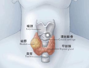
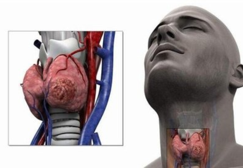
甲状腺癌是发生在甲状腺滤泡或滤泡旁上皮细 胞的恶性肿瘤,是头颈外科常见的恶性肿瘤,发病 率逐年增高,且任何年龄段均可发病 [1-2] ,颈部淋 巴结转移发生较早,而确定是否发生颈部淋巴结转 移对临床手术方案的制订及颈部淋巴结的清扫范围 至关重要 [3-4] ,且清除转移淋巴结与患者的预后相关 [5] 。超声检查是甲状腺癌常用的检查方法,具有 分辨率高、成像清晰、简便易行、无创、无射线、 价格低廉的优势 [6] 。本文探讨高频超声影像学特征 在甲状腺癌颈部淋巴结转移诊断中的影响因素。
1 资料与方法
1.1 一般资料 回顾性分析 2018 年 3 月至 2020 年 2 月本院收治的 96 例甲状腺癌患者的临床资料。 纳入标准:术前完成二维超声和彩色多普勒超声检 查并有清晰的影像学资料;符合手术指征,首次采女 34 例;年龄 24~52 岁, 平均(32.12±3.42)岁;乳头状癌 28 例,滤泡状癌 14 例,髓样癌 10 例,未分化癌 4 例。非转移组:男16 例, 女 24 例; 年龄 22~52 岁, 平 均(31.23±3.25)岁;乳头状癌 20 例, 滤泡状癌 10 例, 髓样癌 7 例, 未分化癌 3 例。两组一般资料比较, 差异无统计学意义(P>0.05),有可比性。
1.2 方法 所有患者术前均进行二维超声和彩色多普勒超声检查。仪器采用彩色超声诊断仪 [ 飞利浦医疗, 国食药监械(准)字 2012 第 3231593 号,型号:G4 xMATRiX iU22],配备 L12-5 探头,频率5~12 MHz。患者取仰卧位, 头部后仰, 可在颈后垫软枕,使颈部充分暴露。按顺序进行甲状腺及双侧颈部淋巴结纵断面、横断面等多个切面的超声扫查。仔细检查甲状腺大小,观察并记录病灶及异常淋巴结超声特征,包括大小、边界、质地回声、位置、微钙化情况、纵横径、距被膜距离(d)、包膜情况、内部血流情况等。
1.3 统计学方法 采用 SPSS 22.0 统计学软件处理数据,计量资料以( ±s )表示,采用 t 检验,计数资料以率(%) 表示, 采用 χ2 检验,多因素分析采用 Logistic 分析,以 P<0.05 为差异有统计学意义。
2 结果
2.1 两组超声影像学特征比较 转移组结节直径≥ 1.5 cm、质地回声不均匀、纵横比≥ 1、微钙化、内部血供丰富、距被膜距离(d)=0 占比均高于非转移组,差异有统计学意义(P<0.05) ;两组边界、形态是否规则占比比较,差异均无统计学意义( P>0.05)。见表 1。
| 表 1 两组超声影像学特征比较 [n (%)] |
| 超声影像特征 |
转移组( n=56) |
非转移组( n=40) |
χ2 值 |
P 值 |
| 结节直径(cm) |
<1.5
≥ 1.5 |
18(32.14)
38(67.86)
26(46.43)
30(53.57)
20(35.71)
36(64.29)
21(37.50)
35(62.50)
23(41.07)
33(58.93)
32(57.14)
24(42.86)
35(62.50)
21(37.50)
31(55.36)
19(33.93)
6(10.71) |
23(57.50)
17(42.50)
21(52.50)
19(47.50)
18(45.00)
22(55.00)
24(60.00)
16(40.00)
33(82.50)
7(17.50)
13(32.50)
27(67.50)
9(22.50)
31(77.50)
7(17.50)
23(57.50)
10(25.00) |
6.132
0.344
0.841
4.744
16.477
5.690
15.038
14.269 |
0.013
0.557
0.359
0.029
0.000
0.017
0.000
0.001 |
| 边界 |
清晰
不清晰 |
|
|
|
|
| 形态 |
规则
不规则 |
|
|
|
|
| 质地回声 |
均匀
不均匀
< 1 |
|
|
|
|
| 纵横比 |
≥1 |
|
|
|
|
| 微钙化 |
有 无 |
|
|
|
|
| 内部血供丰富 |
是
否
d=0 |
|
|
|
|
| 距被膜距离(mm) |
0<d ≤ 2
d>2 |
|
|
|
|
2.2 甲状腺癌淋巴结转移影响因素的多因素分析 以甲状腺癌是否发生颈部淋巴结转移为因变量,将上述 P<0.05 的作为自变量,赋值见表 2。
| 表 2 变量赋值 |
| 因素 |
赋值情况 |
| 结节直径 |
“≥ 1.5 cm”=1,“<1.5 cm”=0 |
| 质地回声 |
“不均匀”=1,“均匀”=0 |
| 纵横比 |
“≥ 1”=1,“<1”=0 |
| 微钙化 |
有 =1,无 =0 |
| 内部血供丰富 |
是 =1,否 =0 |
| 距被膜距离 |
“d=0”=2,“0<d ≤ 2”=1,“d>2”=0 |
Logistic 回归分析结果显示,结节直径 ≥ 1.5 cm、质地回声不均匀、纵横比≥ 1、微钙化、内部血供丰富、距被膜距离(d)=0 等超声检查影像学特征均为甲状腺癌患者发生颈部淋巴结转移的危险因素(P<0.05,OR>1 )。见表3。
表 3 影响甲状腺癌颈部淋巴结转移的多因素分析
| 因素 |
β 值 |
SE |
Wald/χ2 值 |
P 值 |
OR 值 |
95%CI |
| 结节直径≥ 1.5 cm |
1.320 |
0.615 |
4.562 |
0.023 |
3.743 |
1.208~11.599 |
| 质地回声不均匀 |
1.789 |
0.612 |
7.235 |
0.004 |
5.983 |
1.772~20.210 |
| 纵横比≥ 1 |
1.912 |
0.620 |
7.452 |
0.002 |
6.767 |
2.039~22.455 |
| 有微钙化 |
1.742 |
0.534 |
6.452 |
0.001 |
5.540 |
1.945~15.778 |
| 内部血供丰富 |
2.115 |
0.620 |
12.562 |
0.001 |
8.290 |
2.459~27.944 |
| 距被膜距离(d)=0 |
1.745 |
0.875 |
3.875 |
0.046 |
5.726 |
1.030~31.817 |
3 讨论
甲状腺癌的典型特征是甲状腺内出现质地坚硬的肿块, 初诊时部分患者已发生颈部淋巴结转移 [7]。因此,应加强影像学检查,尽早确定是否存在颈部 淋巴结转移,以便制订手术方案和确定淋巴结清扫 范围,降低二次手术率、患者后期的转移率和病死 率。目前,磁共振成像(MRI)、 CT、超声、放射 性核素显像等均是常用的影像学检查方法,但 CT、 放射性核素显像辐射较大, MRI 费用较高、用时较 长,而超声多普勒检查简便、无辐射、无创、价格 低廉,患者易于接受。
本研究结果显示, 转移组结节直径≥ 1.5 cm、 质地回声不均匀、纵横比≥ 1、微钙化、内部血供 丰富、距被膜距离(d)=0 占比均高于非转移组; Logistic 回归分析结果显示,结节直径 ≥ 1.5 cm、 质地回声不均匀、纵横比≥ 1、微钙化、内部血供 丰富、距被膜距离(d)=0 等超声检查影像学特征 均为甲状腺癌患者发生颈部淋巴结转移的危险因素。 分析原因为结节直径≥ 1.5 cm 更易出现颈部淋巴结 转移,可能是因为肿瘤内新生血管丰富加快了结节 生长, 导致结节直径变大, 随着结节生长、增大, 其侵犯周围淋巴管的可能性增大,发生转移的风险 增高 [8] 。同时,癌结节直径越大,其边缘与甲状腺 被膜接触的面积越大,则癌细胞浸润周围淋巴管并 脱落定植生长的风险越大 [9] 。正常甲状腺实质平坦 光滑,超声下回声均匀,若甲状腺发生病变,甲状 腺实质凹凸不平,存在反射点,甲状腺质地回声会 发生改变。已知 90% 的恶性肿瘤超声显示回声不均匀,此时恶性肿瘤生长快,更易侵犯周围淋巴结 而发生转移 [10]。在人体重力作用下, 一般结节横径 大于纵径,但恶性肿瘤会脱离重力作用,向任意方 向生长,可能会导致纵径大于横径。当甲状腺结节 纵横比≥ 1,表明肿瘤细胞浸润导致甲状腺结构被 破坏,纵轴大于横轴,纵横比增加,恶性的可能性 大, 发生转移的风险高 [11]。微钙化与癌细胞快速增 长、结节内血管及纤维组织大量增生,致钙盐沉积 有关;也可能与癌结节自身分泌糖蛋白、黏多糖等 引起钙化有关。而微钙化是甲状腺癌颈部淋巴结转 移的特征性征象,在其他恶性肿瘤中较少出现 [12]。
结节内部血流信号丰富,表明结节内新生血 管丰富,肿瘤细胞生长快,更易发生颈部淋巴结转 移 [13-14] 。而结节边界不清晰,形态不规则是甲状腺 癌浸润性生长的超声特征,表明肿瘤恶性程度和转 移风险均较高 [15]。本研究结果同时显示, 两组边界、 形态是否规则占比比较,差异均无统计学意义。分 析原因可能与本研究纳入的样本量少有关,其结果 尚需后期扩大样本量,开展深入研究予以印证。
综上所述,结节直径≥ 1.5 cm、质地回声不均 匀、纵横比≥ 1、微钙化、内部血供丰富、距被膜 距离(d)=0 等超声检查影像学特征均为甲状腺癌 患者发生颈部淋巴结转移的危险因素。
参考文献
[1] Liu Y, Li B, Zheng Q, et al. The Current Landscape of Clinical Studies Focusing on Thyroid Cancer: A Comprehensive Analysis of Study Characteristics and Their Publication Status[J]. Front Endocrinol, 2020(11): 575799.
[2] 田文,陈志达,郗洪庆,等 . 近红外自体荧光显像技术在甲 状腺癌根治性手术中辅助甲状旁腺识别应用 1 例报告并文献 复习 [J]. 中国实用外科杂志, 2020, 40(5): 591-593.
[3] Joseph FG, Rubtsov D, Davoren P. Appropriateness of ultrasound imaging for thyroid pathology, the standard of radiology reporting on thyroid nodules and the detection rates of thyroid malignancy: a tertiary centre retrospective audit[J]. Intern Med J, 2020, 50(6): 732-740.
[4] Feng Y, Min Y, Chen H, et al. Construction and validation of a nomogram for predicting cervical lymph node metastasis in classic
papillary thyroid carcinoma[J]. J Endocrinol Invest,2021,44(2): 2203-2211.
[5] 周倩,许萍 . 超声评估甲状腺癌颈部转移淋巴结的研究进 展 [J]. 中国医学影像技术, 2019, 35(11): 1752-1756.
[6] 张强,张仑,王旭东,等 . 甲状腺超声智能诊断的现状及研 究进展 [J]. 中国肿瘤临床, 2021, 48(9): 192-196.
[7] 左华,周冬,陈晓军 . 血清 CCL11、GPX3 和 Gal-1 水平在分 化型甲状腺癌诊断中的临床价值 [J]. 检验医学与临床, 2022, 19(5): 586-590.
[8] 毛明锋,杨顺实,刘建新,等 . 超声对甲状腺乳头状癌颈部 转移性淋巴结的预测价值 [J]. 中华实验外科杂志, 2019, 36(7): 1276-1278.
[9]Tao L, Zhou W, Zhan W, et al. Preoperative Prediction of Cervical Lymph Node Metastasis in Papillary Thyroid Carcinoma via Conventional and Contrast-Enhanced Ultrasound[J].
[10] J Ultrasound Med, 2020, 39(10): 2071-2080.Wang M, Li R, Zou X, et al. A miRNA-clinicopathological nomogram for the prediction of central lymph node metastasis in papillary thyroid carcinoma-analysis from TCGA database[J]. Medicine, 2020, 99(35): 21996.
[11]林文金,薛恩生,林振湖,等 . 构建甲状腺癌颈部淋巴结 转移影像报告和数据系统 [J]. 中国医学影像技术, 2021, 37(10): 1452-1455.
关注SCI论文创作发表,寻求SCI论文修改润色、SCI论文代发表等服务支撑,请锁定SCI论文网 文章出自SCI论文网转载请注明出处:https://www.lunwensci.com/yixuelunwen/39342.html