SCI论文(www.lunwensci.com):
摘要:全膝关节置换术已经成为治疗膝关节疾病终末期关节功能障碍的有效的方法,膝关节假体周围感染是该手术灾难性的并发症,不仅增加患者痛苦,而且会导致膝关节功能障碍,甚至面临截肢的风险。尽早诊断和治疗全膝关节术后感染关系到患者的预后。目前对于全膝关节置换术后感染的诊断方法多种多样,各有特点,本文综述了目前对全膝关节置换术后感染的主要诊断方法。
关键词:关节置换;膝关节;感染;诊断
本文引用格式:孙哲,孙朝军,李红,等.初次全膝关节置换术后感染诊断的研究进展[J].世界最新医学信息文摘,2019,19(98):112-114.
Research Progress in the Diagnosis of Infection after Primary Total Knee Arthroplasty
SUN Zhe,SUN Chao-jun,LI Hong,HU Zhi-fu
(The First Affiliated Hospital of Dali University,Dali Yunnan)
ABSTRACT:Total knee arthroplasty has become an effective method to treat the end-stage joint dysfunction of the knee joint disease.The infection around the knee prosthesis is a catastrophic complication of the operation,which not only increases the pain of patients,but also leads to the dysfunction of the knee joint,and even faces the risk of amputation.Early diagnosis and treatment of postoperative infection of total knee joint is related to the prognosis of patients.At present,there are many diagnostic methods for postoperative infection of total knee arthroplasty.
KEY WORDS:Joint replacement;Knee joint;Infection;Diagnosis
0引言
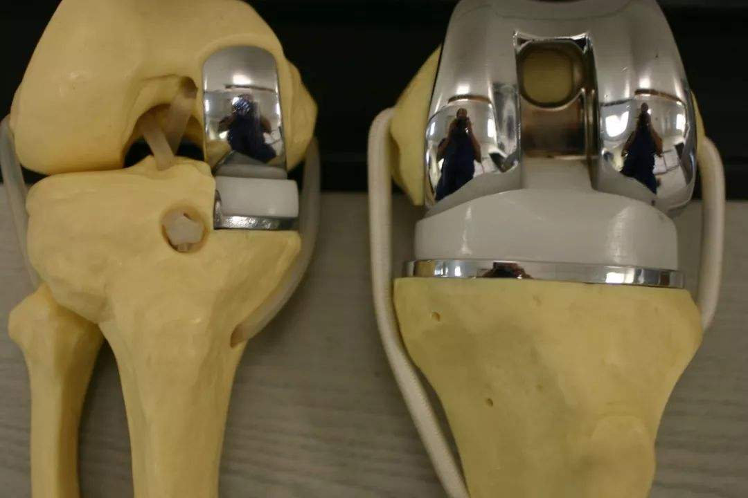
全膝关节置换术(total knee arthroplasty,TKA)是治疗膝关节晚期病变的最有效的手术方法,特别是对于膝关节骨关节炎晚期的患者,全膝关节置换能减轻或消除疼痛,矫正下肢内外翻等畸形,恢复患者的膝关节功能[1-2]。全膝关节置换术的临床应用尽管十分成熟,但是仍然存在术后并发症,术后感染就是最严重并发症。据国外文献报道,初次全膝关节置换术后假体周围感染的发生率为1%~4%[3]。外科医生可以通过术前严格筛选病人,提高手术技术及无菌术水平来降低术后感染率,但每年接受全膝关节置换术的患者越来越多,导致全膝关节置换术后感染的患者也逐年增加。全膝关节置换术后感染的病原菌分布复杂,耐药菌感染比例不断增加[4]。加之,假体周围感染若不早期诊断及治疗,则会产细菌生膜,导致感染更难以控制,给治疗带来极大的困难[5]。膝关节假体周围感染成为膝关节翻修的主要原因,其治疗困难、病死率高,增加患者的医疗负担[6]。所以及时诊断出膝关节假体周围感染十分重要,能减少医疗费用[7],还可以缩短治疗时间,尽早减轻患者痛苦。现就近期全膝关节置换术后感染临床诊断辅助技术新进展作一综述,为临床诊治相关疾病提供参考。
1临床表现
临床工作中,医生必须详细询问、记录患者病史并行体格检查[30]。手术前,询问病史,须特别注意患者是否存在易感因素,包括既往局部及全身感染史、切口周围皮疹、糖尿病史、吸烟史、肥胖、肝功能异常史,免疫疾病史等,这些疾病的存在会增加患者术后假体周围感染的风险。手术后,感染早期病史能为正确的诊断提供重要线索,询问过程中需特别询问患者是否出现发热,并伴有膝关节切口“跳痛”,以及询问患者是否出现“夜间静息痛”,伴有切口周围皮肤红肿,皮温升高等情况。了解手术时间是否延长、术后长时间引流、术口流液等。以上病史询问有助于判断患者是否存在感染症状。体征包括体温、脉搏、血压、呼吸频率。查看患者膝关节周围是否出现红、肿、热、痛,主被动活动受限,膝关节腔积液是否逐渐增加,出现以上体征往往提示可能存在假体周围感染,如果发现手术侧膝关节切口周围与假体相通的窦道则可以直接确诊假体感染。但是有些亚急性、慢性或者低毒性感染者临床表现不明显,根据病史和症状不能确诊,因此需要结合实验室检查来辅助诊断。
2血清学检查
反应蛋白(C-reactive protein,CRP)是在机体受到感染或组织损伤时血浆中一些急剧上升的蛋白质(急性蛋白),激活补体和加强吞噬细胞的吞噬而起调理作用,清除入侵机体的病原微生物和损伤,坏死,凋亡的组织细胞。红细胞沉降率(erythrocyte sedimentation rate,ESR)是以红细胞在第一小时末下沉的距离来表示红细胞的沉降速度,简称血沉。C反应蛋白(CRP)和红细胞沉降率(ESR)是诊断全膝关节置换术后的最常用指标。关节置换术后CRP于2-3 d达到高峰,14-21 d回到正常范围[14-15]。Greidanue等[9]发现,关节置换后ESR于5-7 d达到高峰,3个月到1年时间内回到正常范围内。一项对2736名患者的数据进行的meta分析显示,对于关节置换术后假体周围感染,ESR的敏感性和特异性分别为86%和72.3%,CRP水平分别为86.9%和78.6%[8]。如果血沉和CRP结果都在正常水平内,关节置换术后感染的可能性很低[9]。但是,Mcarthur BA[10]等指出,约有4%的假体周围感染者ESR和CRP均正常。所以,诊断膝关节置换术后假体周围感染需要结合病史、体征及实验室检查,必要时结合关节腔穿刺。
IL-6是是全身炎症反应综合征的敏感指标[11],近年来,逐渐应用于感染筛查[12]。有研究认为[13],在关节置换术后IL-6可在30-430 pg/ml范围波动,2天内达到高峰并下降。Di Cesare等[12]认为IL-6用于诊断膝关节感染,其敏感性是100%,特异性是95%。关节置换术后2天内怀疑感染可以使用IL-6检测,为诊断感染早期提供依据。近年来,有学者已经证明血清降钙素原(PCT)能提高对细菌感染和脓毒血症的诊断率[16]。闫立伟等[17]的研究证实了PCT在诊断膝关节置换术后早期感染具有重要临床价值。Yuan等[18]研究发现,当降钙素原>0.5 ng/mL时,其诊断假体感染的敏感性为80%,特异性为73.9%。
3关节液分析
白细胞酯酶是由感染部位激活的中性粒细胞产生的酶,最初被用于协助诊断尿路感染[19],关节液白细胞酯酶测试是一种快速、廉价、微创的试验方法[20],可以早期对关节假体周围感染进行诊断。有文献研究报道,以(2+)读数作为关节置换假体周围感染的临界值,敏感性为80.6%。白细胞酯酶测试与血清ESR及CRP,关节液白细胞计数和中性细胞百分比比较,对假体周围感染的诊断有更好的价值[21]。一项meta分析显示白细胞酯酶诊断假体周围感染的敏感性为77%,特异性为95%,但是关节液混有血液时,可影响试纸条结果[22]。
α-防御素由活化的中性粒细胞释放,是宿主内具有广谱抗菌性的活性小分子肽类,对革兰氏阳性、阴性菌均具有广谱抗菌活性。李睿,陈继营[23]认为,α-防御素是,诊断人工关节置换术后假体周围感染的理想标记物。Frangiamore SJ等[24]研究报道,α-防御素以5.2mg/L为诊断界值时,其诊断假体周围感染敏感性为100%,特异性为98%,阳性预测值为96%,阴性预测值为100%。有研究表明[25-26],α-防御素的高特异性不受致病菌类型及毒力的影响,也不受患者合并有系统性炎性疾病或使用抗生素的影响。
近年来有学者报道关节液CRP对关节置术后感染有很高的价值[27]。Lee等[22]研究报道,关节液CRP诊断假体周围感染的敏感性为85%,特异性为88%。关节液CRP对于诊断假体周围感染具有更高的敏感性和特异性,关节液CRP>2.78mg/L时,诊断感染的敏感性为100%,特异性为82%,关节液CRP>5.37mg/L,诊断感染的敏感性为90%,特异性将提升至91%[28]。
膝关节穿刺液白细胞计数和中性粒细胞百分比容易获取、便于计数,具有良好的诊断价值。时间在术后6周内,白细胞计数大于10000个/μL,关节液中性粒细胞百分比大于90%考虑假体周围感染。时间在术后6周之后,白细胞计数大于3000个/μL,关节液中性粒细胞百分比大于80%考虑假体周围感染[29]。关节液白细胞计数及中性粒细胞百分比诊断膝关节置换假体周围感染敏感性及特异性分别是,关节液白细胞计数敏感性为89%,特异性为86%,中性粒细胞百分比敏感性89%,特异性86%[22]。
关节液细菌培养是2018年人工关节置换术后假体周围感染诊断新定义中的重要标准[31]。细菌培养诊断特异性高,但培养结果阴性不能排除假体周围感染[32]。
抗生素的使用会导致培养结果阴性,故建议停用抗生素2周以上再行关节液穿刺培养[33-34]。也有学者建议,在穿刺前需停抗生素药2~3周,并且将关节液培养时间延长至5~10天增加培养阳性率[35]。使用血培养瓶培养对假体周围感染诊断的敏感性和特异性分别为92.1%和99.7%,并且使用需氧和厌氧的培养血培养瓶,其诊断敏感性与特异性均高于单纯使用血培养瓶培养[36]。
4病理学检查
对于一些未能通过非手术方式确诊的怀疑假体周围感染的且接受再次手术的患者,手术过程中应取假体周围组织做冰冻切片行病理学检查,同时取假体周围炎性组织行组织细菌培养[33]。Parvizi J等[37],建议至少取3块假体周围组织行术中冰冻切片,每个高倍视野出现多于5个白细胞为阳性结果。Mirra等[38]认为冰冻组织切片在400倍的高倍镜视野中,至少3个高倍视野中每个视野可见≥5个中性白细胞考虑感染。Kwiecien G等[39]研究发现,冰冻切片的敏感性、特异性分别为73.7%、98.8%。Tsaras G[40]和Wu C[41]也认为术中冷冻病理切片检查在诊断假体周围感染方面特异性较高,当其他检验结果不能确诊感染时,其具有良好的应用价值。但这项检查同时依赖外科医生和病理科医生的丰富经验。
5影像学检查
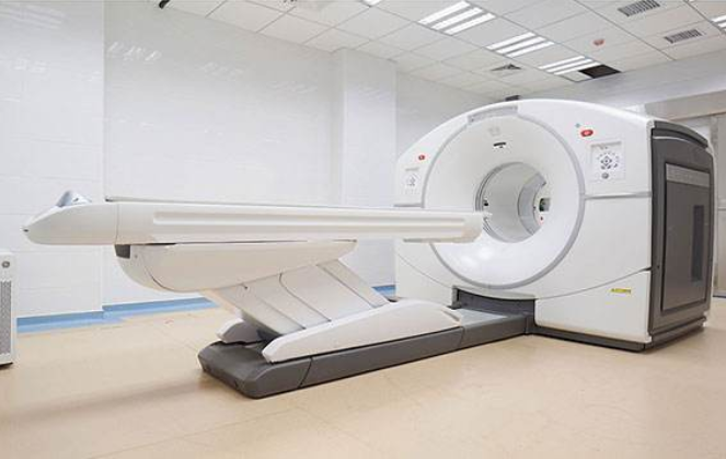
X线平片在感染发生3~6个月后,X线影像才会出现改变,动态连续监测可以作为区分感染性松动和机械性松动的标志[42]。CT与MRI对于钛或钽以外的假体在成像上会出现干扰伪影,在诊断假体周围感染时也缺乏特异性,不常规用于诊断假体感染。核医学为假体周围感染诊断带来新的思路。还有学者提出[33],可标记白细胞成像与骨或骨髓成像来诊断假体周围感染,如,F-18氟脱氧葡萄糖-正电子发射断层扫描(FDG-PET)成像,镓成像或标记白细胞成像等。Trevail C等[43]研究发现,通过99m锝骨扫描及111铟标记白细胞扫描结合骨髓扫描可以使假体周围感染的诊断准确率到达为97.7%,特异性达到99.5%,敏感性达到80%。
6分子生物学技术
近年来,聚合酶链反应(PCR)技术成为诊断假体周围感染的热门方法。其中细菌共有基因16S rRNA是微生物诊断的研究热点,文献[44]对115例人工关节置换失败患者的关节液标本进行以16S rRNA作为引物的PCR分析,实验结果为PCR组诊断假体周围感染的敏感性为71%,而常规培养组为44%。Trampuz A等[45]认为,PCR技术可提高关节假体周围感染的细菌检出率,特别是对于培养阴性的标本。Birmingham P[46]认为PCR技术虽然敏感性高,但在细菌被抗生素清除死亡时会表现出假阳性。因此,有学者[47]使用16S rRNA逆转录PCR诊断假体周围感染,得到诊断感染的敏感性为71%,特异性为100%。因为逆转录PCR以RNA为模板,RNA有着被分离出来后易降解的特性,这使检测结果不易受到样本污染干扰,结果更加准确[46,47]。但是PCR技术的主要问题还是在于较高的假阳性率,导致该方法目前仍难以推广[48]。
7结论与展望
综上所述,全膝关节置换术后感染是较严重的并发症,目前所有辅助检查方法都存在各自的敏感性或者特异性,因此每一种方法都存在一定局限性。笔者认为,对于诊断关节假体周围感染,仔细询问患者病史与体格检查仍然是一种最重要和最简便可行的初始筛查方法。所有假体周围感染的可疑者须行ESR、CRP检测,若检查结果正常,而患者又出现膝关节切口周围肿胀及夜间疼痛加重或者不缓解,这时需要行关节穿刺与滑液分析,并且送关节液需氧和厌氧血培养,若患者存在发热时,需在体温最高处,连续抽血培养检查。普通影像学诊断假体周围感染能力有限,可考虑应用核医学成像技术辅助诊断。对于不能用非手术方式明确诊断的可疑患者,若行手术,则术中必须行冰冻切片病理检查有利于明确诊断和指导下一步治疗。新型滑液标记物及分子生物学技术的应用为假体周围感染诊断提供了新的方向,有望为关节置换假体周围感染诊断提供新的方法。
参考文献
[1]Arden NK,Leyland KM.Osteoarthritis year 2013 in review:clinical[J].Os-teoarthritis Cartilage,2013,21(10):1409-1413.
[2]Dziedzic KS,Healey EL,Porcheret M,et al.Implementing the NICE os-teoarthritis guidelines:a mixed methods study and cluster randomised trial of a model osteoarthritis consultation in primary care-the Management of Os-teoArthritis In Consultations(MOSAICS)study protocol[J].Implement Sci,2014,9:95.
[3]Kim YH,Park,JW,Kim JS,et al.The outcome of infented total knee arthrop-lasty:culture-positive versus culture-negative[J].Arch Orthop Traum Su,2015,135(10):132-139.
[4]AggarwalnVK,Bakhshi H,Ecker NU,et al.Organism profile in periprosthetic joint infection:pathogens differ at two arthroplasty infection referral centers in Europe and in the United States[J].J Knee Surg,2014,27(5):399-406.
[5]Gbejuade HO,Lovering AM,Webb JC.The role of microbial biofilms in prosthetic joint infections[J].Acta Orthop,2015,86(2):147-158.
[6]Springer BD.The diagnosis of periprosthetic joint infection[J].J Arthroplas-ty,2015,30(6):908-911.
[7]Zimmerli W,Trampuz A.Prosthetic joint infections:update in diagnosis and treatment[J].Swiss Med Wk,2005,13(2):243.
[8]Huerfano E,Bautista M,Huerfano M.Screening for Infection Before Revision Hip Arthroplasty:A Meta-analysis of Likelihood Ratios of Erythrocyte Sedi-mentation Rate and Serum C-reactive Protein Levels[J].J Am Acad Orthop Surg,2017,25(12):809-817.
[9]Greidanus NV,Masri BA,Garbuz DS,et al.Use of erythrocyte sedimentation rate and C-reactive protein level to diagnose Infection before revision total knee arthroplasty.A prospective evaluation[J].J Bone Joint Surg Am,2007,89(7):1 409-1 416.
[10]Mcarthur BA,Abdel MP,Taunton MJ,Et al.Seronegative infections in hip and knee arthroplasty:periprosthetic infections with normal erythrocyte sedimen-tation rate and C-reactive protein level[J].Bone Joint J,2015,97-B(7):939-944.
[11]Berbari E,Mabry T,Tsaras G,et al.Inflammatory blood laboratory levels as markers of prosthetic joint infection:a systematic review and meta-analysis[J].Bone Joint Surg Am,2010,92(11):2102-2109.
[12]Di Cesare PE,Chang E,Preston CF,et a1.Serum interleukin-6 as a marker of periprosthetic infection following total hip and knee arthroplasty[J].JBone Joint Surg Am,2005,87(9):1921-1927.
[13]Bottnee F,Wevnvr A,Winkelmann W,et al.Interleukin-6,procalcitonin and TNF-alpha:markers of periprosthetic infection following total joint replace-ment[J].J Bone Joint Surg Be,2007,89:94-96.
[14]王岩,郝立波,周勇刚,等.人工髋关节置换术后的临床经验分析[J].中华外科杂志,2005,43:1313-1316.
[15]Parvizi J,Ghanem E,Menashe S.Periprosthetic infection:what are the diag-nostic challenges?[J].J Bone Joint Surg Am,2006,88:138-147.
[16]Hunziker S,Hugle T,Schuchardt K,et al.The value of serum procalcitonin level for differentiation of infectious from noninfectious causes of fever after orthopaedic surgery[J].J Bone Joint Surg Am,2010,92(1):138-148.
[17]闫立伟,王文波.李昭旭,等.血清降钙素原在诊断膝关节置换术后早期感染的意义[J].中国骨与关节杂志,2015,14(4):317-319.
[18]Yuan K,Li WD,Qiang Y,et al.Comparison of procalcitonin and C-reactive protein for the diagnosis of periprosthetic joint infection before revision total hip arthroplasty[J].Surg Infect(Larchmt),2015,16(2):146-150.
[19]Rowe TA,Juthani-Mehta M.Diagnosis and management of urinary tract infection in older adults[J].Infect Dis Clin North Am,2014,28(1):75-89.
[20]Parvizi J,Jacovides C,Antoci V,et al.Diagnosis of periprosthetic joint infec-tion:the utility of a simple yet unappreciated enzyme[J].J Bone Joint Surg Am,2011,93(24):2242-2248.
[21]Shahi A,Tan TL,Kheir MM,et al.Diagnosing periprosthetic joint infec-tion:and the winner is?[J].J Arthroplasty,2017,32(9S):S232-S235.
[22]Lee YS,Koo KH,Kim HJ,et al.Synovial fluid biomarkers for the diagnosis of periprosthetic joint infection:a systematic review and meta-analysis[J].J Bone Joint Surg Am,2017,99(24):2077-2084.
[23]李睿,陈继营.人工关节置换术后假体周围感染诊断方法的研究进展[J].中华骨科杂志,2016,36(19):1254-1262.
[24]Frangiamore SJ,Gajewski ND,Saleh A,et al.α-defensin accuracy to diagnose periprosthetic joint infection-best available test?[J].J Arthroplasty,2016,31(2):456-460.
[25]Deirmengian C,Kardos K,Kilmartin P,et al.The alpha-defensin test for peri-prosthetic joint infection responds to a wide spectrum of organisms[J].Clin Orthop Relat Res,2015,473(7):2229-2235.
[26] Deirmengian C,Kardos K,Kilmartin P,et al.The alpha-defensin test for periprosthetic joint infection outperforms the leukocyte esterase test strip[J].Clin Orthop Relat Res,2015,473(1):198-203.
[27]Parvizi J,Jacovides C,Adeli B,et al.Coventry Award:synovial C-reactive pro-tein:a prospective evaluation of a molecular marker for periprosthetic knee joint infection[J].Clin Orthop Relat Res,2012,470(1):54-60.
[28]Ronde-Oustau C,Diesinger Y,Jenny JY,et al.Diagnostic accuracy of in-tra-articular C-reactive protein assay in periprosthetic knee joint infection a preliminary study[J].Orthop Traumatol Surg,2014,100(2):217-220.
[29]Parvizi J,Gehrke T,Chen AF.Proceedings of the International Consensus on Periprosthetic Joint Infection[J].Bone Jt J,2013,95-B(11):1450-1452.
[30]Della VC,Parvizi J,Bauer TW,et al.Diagnosis of periprosthetic joint infections of the hip and knee[J].J Am Acad Orthop Surg,2010,18(12):760-770.
[31]Parvizi J,Tan TL,Goswami K,et al.The 2018 Definition of periprosthetic hip and knee infection:an evidence-based and validated criteria[J].J Arthroplas-ty,2018,33(5):1309-1314.
[32]王悦妮,赵秀丽.人工关节假体感染的细菌培养[J].中国组织工程研究,2012,16(22):4129-4132.
[33]Della VC,Parvizi J,Bauer TW,et al.Diagnosis of periprosthetic joint infections of the hip and knee[J].J Am Acad Orthop Surg,2010,18(12):760-770.
[34]Parvizi J,Della VCJ.AAOS Clinical Practice Guideline:diagnosis and treat-ment of periprosthetic joint infections of the hip and knee[J].J Am Acad Or-thop Surg,2010,18(12):771-772.
[35]付玉娟,张超远,乔吉明.人工膝关节置换术后感染误诊l8例分析[J].中国误诊学杂志,2005,28(2):329.
[36]Peel TN,Dylla BL,Hughes JG,et al.Improved diagnosis of prosthetic joint infection by culturing periprosthetic tissue specimens in blood culture bottles[J].MBio,2016,7(1):e01715-e01776.
[37]Parvizi J,Gehrke T.Definition of periprosthetic joint infection[J].J Arthrop-lasty,2014,29(7):1331.
[38]Mirra JM,Marder RA,Amstutz HC.The pathology of failed total joint arth-roplasty[J].Clin Orthop Relat Res,1982,(170):175-183.
[39]Kwiecien G,George J,Klika AK,et al.Intraoperative frozen section histolo-gy:matched for musculoskeletal infection society criteria[J].J Arthroplas-ty,2017,32(1):223-227.
[40]Tsaras G,Madukaezeh A,Inwards CY,et al.Utility of intraoperative frozen section histopathology in the diagnosis of periprosthetic joint infection:a sys-tematic review and meta-analysis[J].J Bone Joint Surg Am,2012,94(18):1700-1711.
[41]Wu C,Qu X,Mao Y,et al.Utility of intraoperative frozen section in the diag-nosis of periprosthetic joint infection[J].PLoS One,2014,9(7):e102346.
[42]Esposito S,Leone S.Prosthetic joint infections:microbiology,diagno-sis,management and prevention[J].Int J Antimicrobi Agents,2008,32(4):287.
[43]Trevail C,Ravindranath-Reddy P,Sulkin T,et al.An evaluation of the role of nuclear medicine imaging in the diagnosis of periprosthetic infections of the hip[J].Clin Radiol,2016,71(3):211-219.
[44]Gallo J,Kolar M,Dendis M,et al.Culture and PCR analysis of joint fluid in the diagnosis of prosthetic joint infection[J].New Microbiol,2008,3l(1):97-104.
[45]Trampuz A,Osmon DR,Hanssen AD,et al.Molecular and antibiofilm ap-proaches to prosthetic joint infection[J].Clin Orthop Relat Res,2003(414):69-88.
[46]Birmingham P,Helm JM,Manner PA,et a1.Simulated joint infection assess-ment by rapid detection of live bacteria with real time reverse transcription polymerase chain reaction[J].J Bone Joint Surg Am,2008,90(3):602-608.
[47]Bergin PF,Doppelt JD,Hamilton WG,et a1.Detection of periprosthetic infections with use of ribosomal RNA-based polymerase chain reaction[J].J Bone Joint Surg Am,2010,92(3):654-663.
[48]Panousis K,Grigoris P,Butcher I,et a1.Poor predictive value of broad-range PCR for the detection of arthroplasty infection in 92cases[J].Acta Or-thop,2005,76(3):341-346.
关注SCI论文创作发表,寻求SCI论文修改润色、SCI论文代发表等服务支撑,请锁定SCI论文网! 文章出自SCI论文网转载请注明出处:https://www.lunwensci.com/yixuelunwen/25633.html


