


SCI论文(www.lunwensci.com)
【摘要】 目的: 分析弥散加权成像(DWI)在宫颈癌诊断中的效能。方法: 选取 2020 年 7 月至 2022 年 5 月该院收治的 96 例疑似 宫颈癌患者为研究对象,均于术前进行磁共振成像(MRI)平扫和 DWI 检查,以术后病理结果为金标准,比较 MRI 平扫、DWI 检查对宫 颈癌的检出结果及在宫颈癌诊断中的效能。结果: 病理学检查结果显示,96 例疑似宫颈癌患者中阳性 65 例,阴性 31 例(其中宫颈糜烂 12 例,子宫肌瘤 19 例) ;MRI 平扫检出阳性 51 例,阴性 45 例;DWI 检查检出阳性 64 例,阴性 32 例;DWI 检查诊断宫颈癌的灵敏度、 准确度、阴性预测值均高于 MRI 平扫,漏诊率低于 MRI 平扫,差异有统计学意义(r<0.05)。 结论: DWI 检查诊断宫颈癌的效能高于 MRI 平扫。
【关键词】 宫颈癌;磁共振成像平扫;弥散加权成像;诊断;效能
Efficiency of diffusion weighted imaging in diagnosis of cervical cancer
【 Abstract 】 Objective: To analyze efficiency of diffusion weighted imaging (DWI) in diagnosis of cervical cancer. Methods: 96 patients with suspected cervical cancer admitted to the hospital from July 2020 to May 2022 were selected as the research objects. All patients underwent magnetic resonance imaging (MRI) plain scan and DWI examination before operation. Using the the postoperative pathological results as the gold standard, the detection results of MRI plain scan and DWI examination for cervical cancer and their efficiencies in the diagnosis of cervical cancer were compared. Results: The pathological examination showed that 65 of the 96 patients with suspected cervical cancer were positive and 31 were negative (including 12 cases of cervical erosion and 19 cases of uterine fibroids)。 MRI plain scan detected 51 positive cases and 45 negative cases. DWI examination detected 64 positive cases and 32 negative cases. The sensitivity, the accuracy and the negative predictive value of DWI examination in the diagnosis of cervical cancer were higher than those of MRI plain scan, the missed diagnosis rate was lower than that of MRI plain scan, and the differences were statistically significant (r<0.05)。 Conclusions: The efficiency of DWI examination in the diagnosis of cervical cancer is higher than that of MRI plain scan.
【Keywords】 Cervical cancer; Magnetic resonance imaging plain scan; Diffusion weighted imaging; Diagnosis; Efficiency
宫颈癌是妇科常见恶性肿瘤,其发病趋于年 轻化,且病死率较高,严重威胁患者生命安全 [1-2]。 因此早期诊断宫颈癌,对制订诊疗方案、提高患者 生存率十分关键 [3-4] 。磁共振成像(MRI)平扫可 了解肿瘤大小、侵犯程度及淋巴结转移情况,但其 扫描时间较长,判断肿瘤分期的准确性不高。弥散 加权成像(DWI)可检测活体组织内水分子的弥散 运动,反映肿瘤的形态学结构和功能信息,利于宫 颈癌的早期诊断 [5-6] 。本文分析 DWI 检查在宫颈癌 诊断中的效能。
1 资料与方法
1.1 一般资料 选取 2020 年 7 月至 2022 年 5 月 本院收治的 96 例疑似宫颈癌患者为研究对象。纳入标准:存在不规则阴道出血或阴道排液等症状;
未接受过放化疗;无 MRI、DWI 检查禁忌证。排除标准:合并心、肾等重要脏器功能障碍;妊娠或哺乳期;合并精神异常。其中年龄 35——71 岁,平均(51.32±4.72) 岁; 症状出现时间 2——20 个月, 平均(11.03±4.26) 个月。
1.2 方法 所有研究对象均于术前进行 MRI 平扫和 DWI 检查。
MRI 平扫:嘱患者检查前禁食 5——6 h, 并适量饮水保持膀胱充盈。采用 Fophie Free 型磁共振成像系统(洛阳康达卡乐福医疗科技有限公司,国械注准 20233061385) 及配套梯度线圈,检查前嘱患者取下身上金属物品,取仰卧位,从耻骨联合至肾门水平进行常规横断位、冠状位和矢状位平扫。设置T1WI 扫描参数:TR 500 ms,TE 15 ms;T2WI 扫描参数: TR 600 ms,TE 60 ms。层厚 4.0 mm, 层距1.0 mm, 视野 300 mm ×250 mm, 矩阵 256×256。
MRI 平 扫 结 束 后 行 DWI 检 查。 设 置 DWI 扫 描参数:层厚 4.0 mm,层距 1.0 mm,TR 5600 ms, TE 83.5 ms ,b 值 800 s/mm2 ,脉冲重复继发次数 2 次。
所有图像均由 2 名 5 年以上资历的影像学医师 进行双盲评判,若意见出现分歧,则由第 3 位副主 任医师进行评判。
1.3 观察指标 (1)术后均行病理学检查,统计 病理学检查结果。(2)以术后病理结果为金标准, 比较 MRI 平扫、DWI 检查对宫颈癌的检出结果。 (3)分析 DWI 检查在宫颈癌诊断中的效能。灵敏度 = 真阳例数 /(真阳 + 假阴)例数 ×100%; 特异度 = 真阴例数 /(假阳 + 真阴)例数 ×100%; 准确度 = (真阳 + 真阴) 例数 / 总例数 ×100%。阳性预测值 = 真阳例数 /(真阳 + 假阳) 例数 ×100%; 阴性预测 值 = 真阴例数 /(假阴 + 真阴)例数 ×100%。漏 诊率 =1- 灵敏度;误诊率 =1- 特异度。
1.4 统计学方法 采用 SPSS 22.0 统计学软件处理 数据,计数资料以率(%)表示,采用 χ2 检验,以 P<0.05 为差异有统计学意义。
2 结果
2.1 病理学检查结果 病理学检查结果显示,96 例疑似宫颈癌患者中阳性 65 例,阴性 31 例(其中 宫颈糜烂 12 例,子宫肌瘤 19 例)。
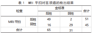
2.2 MRI 平 扫、DWI 检 查 对 宫 颈 癌 的 检 出 结 果 96 例疑似宫颈癌患者中,MRI 平扫检出阳性 51 例, 阴性 45 例; DWI 检查检出阳性 64 例,阴 性 32 例。见表 1、表 2。

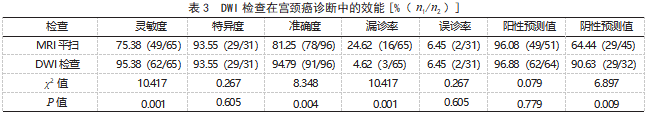
2.3 DWI 检查在宫颈癌诊断中的效能 DWI 检查诊断宫颈癌的灵敏度、准确度、阴性预测值均高于MRI 平扫,漏诊率低于 MRI 平扫,差异有统计学意义(P<0.05) 。见表 3。
3 讨论
宫颈癌的发生与人乳头瘤病毒感染、吸烟等因素有关 [7] 。患者早期常无明显症状和体征,极易发生漏诊、误诊,导致疾病早期诊断困难 [8-9]。
近年来,影像学检查已成为诊断宫颈癌的重要手段, MRI 平扫对软组织的分辨率较高,可反映病灶部位、大小、信号特点及浸润情况 [10-11] ,其应用于宫颈癌患者,可表现为类圆形或不规则肿块,在T2WI 上表现为均匀或欠均匀的高信号,与正常宫颈组织形成良好对比 [12]。
DWI 是目前唯一能在活体观察组织水分子微观运动的无创性影像学方法,可有效检测与组织含水量变化相关的形态学和生理学早期改变,并以表观弥散系数(ADC)值来量化表示,可清晰反映肿瘤病灶及其临床分期 [13-14]。
本研究结果显示, 病理学检查结果显示, 96 例疑似宫颈癌患者中阳性 65 例, 阴性 31 例; MRI 平扫检出阳性 51 例,阴性 45 例;DWI 检查检出阳性64 例, 阴性 32例;且 DWI 检查诊断宫颈癌的灵敏度、准确度、阴性预测值均高于 MRI 平扫,漏诊率低于 MRI 平扫。分析原因为 MRI 平扫通过使子宫基层、宫颈基质及宫颈癌病灶周围组织对比度的差异来获得强化图像,从而显示病灶部位,但其诊断准确率有待提高 [15-16] 。已知当组织含水量丰富、微循环灌注量较高或细胞外间隙较大时,水分子扩散受限程度轻,ADC 值较大 [17-18] ;而组织内细 胞及间质成分较多、细胞外间隙小时,ADC 值较 小 [19-20] 。宫颈癌细胞分裂活跃、生长迅速,细胞排 列密集、间隙小,且肿瘤细胞内液较少,水分子在 细胞内外的扩散速率低,DWI 检查时显示为 ADC 值变小,因此能够有效区分癌灶组织与正常宫颈组 织 [21-22]。因此, DWI 检查用于诊断宫颈癌可提高灵 敏度、准确度,降低漏诊率。
综上所述,DWI 检查诊断宫颈癌的效能高于 MRI 平扫。
参考文献
[1] 王贝贝,梁慧,李观得,等 . 单核苷酸多态性与宫颈癌患者 易感性的相关研究 [J]. 武警后勤学院学报(医学版) ,2021, 30(7): 91-92.
[2] 陈鸿雁 . 磁共振弥散加权成像在宫颈癌放疗疗效和预后评估 中的应用价值及符合率评价 [J]. 现代医用影像学,2022,31 (11):2077-2079.
[3] Miao L ,Cao Y ,Zuo L ,et al. Predicting pathological complete response of neoadjuvant radiotherapy and targeted therapy for soft tissue sarcoma by whole-tumor texture analysis of multisequence MRI imaging[J]. Eur Radiol ,2023 ,33(6): 3984-3994.
[4] 李刚,祝青,蒲红波,等 . 1.5T 磁共振 DWI 序列在宫颈癌诊 断及临床分期中的价值分析 [J]. 中国 CT 和 MRI 杂志, 2020, 18(9): 123-125.
[5] 黎政华,岑贤友,王治 . 磁共振多 b 值弥散加权成像对宫 颈癌宫旁浸润的诊断价值 [J]. 医疗装备,2022,35(16): 42-44.
[6] Huang Q ,Deng B ,Wang Y ,et al. Reduced field-of-view DWI-derived clinical-radiomics model for the prediction of stage in cervical cancer[J]. Insights Imaging ,2023 ,14(1): 18.
[7] 张春林,龚权,梅冰 . Toll 样受体在人乳头瘤病毒感染致宫颈 癌中的作用 [J]. 中华微生物学和免疫学杂志,2022,42(1) : 73-77.
[8] 李长海 . 磁共振成像平扫及弥散加权成像在宫颈癌术前分期诊 断中的应用价值 [J]. 河南医学研究,2023,32(5):930-932.
[9] 李小燕,孙洪赞,卢再鸣 . 宫颈癌宫旁浸润影像诊断研究进 展 [J]. 现代肿瘤医学, 2020 ,28(9): 1571-1574.
[10] Cai SQ ,Song ZY ,Wu MR ,et al. Magnetic resonance imaging and diffusion weighted imaging-based histogram in predicting mesenchymal transition high-grade serous ovarian cancer[J]. Acad Radiol ,2023 ,30(6): 1118-1128.
[11] Qin Y ,Tang C ,Hu Q ,et al. Quantitative assessment of restriction spectrum MR imaging for the diagnosis of breast cancer and association with prognostic factors[J]. J Magn Reson Imaging,2023 ,57(6): 1832-1841.
[12] Holopainen E ,Lahtinen O ,K。n。nen M ,et al. Greater increases in intratumoral apparent diffusion coefficients after chemoradiotherapy predict better overall survival of patients with cervical cancer[J]. PLoS One ,2023 ,18(5): 5786.
[13] Xu Q ,Xu Y ,Wang J ,et al. Distinguishing mesorectal tumor deposits from metastatic lymph nodes by using diffusion-weighted and dynamic contrast-enhanced magnetic resonance imaging in rectal cancer[J]. Eur Radiol ,2023 ,33(6): 4127-4137.
[14] Meng T ,Liu H ,Liu J ,et al. The investigation of reduced field of-view diffusion-weighted imaging ( DWI ) in patients with nasopharyngeal carcinoma: comparison with conventional DWI[J].Acta Radiol ,2023 ,64(6): 2118-2125.
[15] 杨涛,程敬亮,王伟,等 . DWI 联合 MRI 常规序列对宫颈癌的诊断及分期与病理对照分析 [J]. 医学影像学杂志,2016,26(7): 1275-1277.
[16] Jung HN ,Ryoo I ,Suh S ,et al. Evaluating the elasticity of metastatic cervical lymph nodes in head and neck squamous cell carcinoma patients using DWI-based virtual MR elastography[J].Magn Reson Med Sci ,2022,16:123-125.
[17] 林会娟,刘海燕,李绍东,等 . 磁共振成像增强扫描联合不同 b 值弥散加权成像在宫颈癌术前分期的诊断价值研究 [J].中国临床医生杂志,2023,51(12):1480-1484.
[18] Jacobsen MC,Rigaud B,Simiele SJ, et al. Feasibility of quantitative diffusion-weighted imaging during intra-procedural MRI-guided brachytherapy of locally advanced cervical and vaginal cancers[J]. Brachytherapy,2023,22(6):736-745.
[19] He Y,Wang M,Yi S, et al. Diffusion-weighted imaging in the assessment of cervical cancer:comparison of reduced field-of-view diffusion-weighted imaging and conventional techniques[J].Acta Radiol,2023,64(8):2485-2491.
[20] 李 媛 媛, 何 源 青 . DWI 联 合 DCE-MRI 在 宫 颈 癌 诊 断 及 分期、淋巴结转移评估中的应用价值 [J]. 临床医学研究与实践,2023,8(19):116-119.
[21] 崔岩 . MRI 平扫联合 DWI 检查在宫颈癌病理分期诊断中的应用价值 [J]. 哈尔滨医药,2023,43(3):100-102.
[22] 王雯智,石建勇,郑蕾,等 . IVIM-DWI 参数联合血清肿瘤标志物检测在宫颈癌诊断及分化程度评估中的应用价值 [J]. 中国 CT 和 MRI 杂志,2023,21(12):129-131.
关注SCI论文创作发表,寻求SCI论文修改润色、SCI论文代发表等服务支撑,请锁定SCI论文网!

本次研究将我院于 2016 年 6 月至 2017 ... 详细>>

如何设计有效的环境治理政策, 是学术界和政策... 详细>>
